

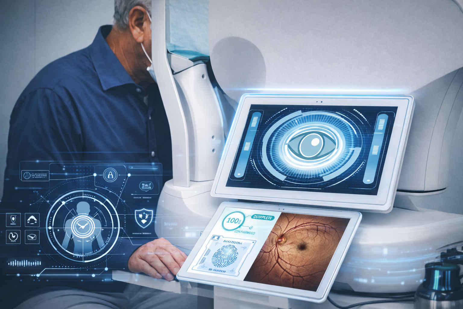
Comprehensive Eye Exams
A comprehensive eye exam evaluates the overall health of your eyes and vision using several advanced diagnostic tools. During the exam, doctors may use Optos retinal imaging to capture a wide view of the back of the eye, helping detect early signs of retinal disease. Optical Coherence Tomography (OCT) provides detailed cross-section images of the retina, allowing doctors to examine its layers and identify conditions such as macular degeneration or glaucoma. A visual field test measures your peripheral vision and helps detect vision loss that may be linked to glaucoma or nerve damage. Together, these technologies give doctors a clearer and more complete understanding of your eye health, allowing problems to be detected earlier and treated more effectively.
Optos is a modern eye scan that lets doctors take a very wide picture of the back of your eye, called the retina. It can capture up to 200 degrees of the retina in one quick image, which is much more than a regular eye exam can see. The scan only takes a few seconds, is non-invasive, and usually doesn’t require dilating your pupils, so you don’t have to deal with blurry vision afterward. Because the image shows such a large area of the retina, doctors can more easily spot early signs of problems like retinal tears, glaucoma, macular degeneration, or diabetic eye disease. The images are saved digitally so your doctor can compare them over time and monitor any changes in your eye health.
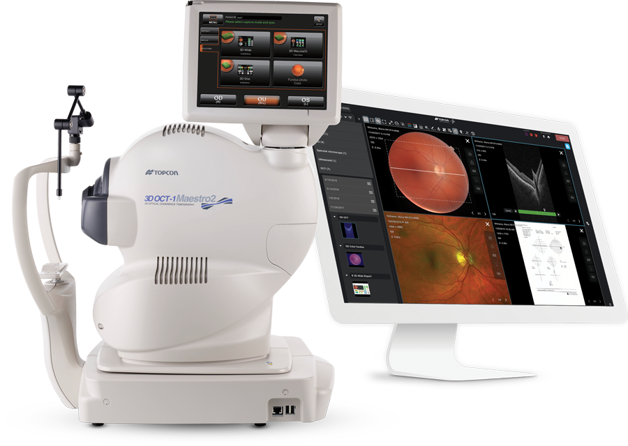
Optical Coherence Tomography (OCT) is an advanced imaging test that allows doctors to see detailed cross-section images of the retina. Using light waves, the scan creates highly precise pictures of the different layers of the retina, helping detect problems that may not be visible during a regular eye exam. The test is quick, painless, and completely non-invasive. OCT helps doctors diagnose and monitor conditions such as glaucoma, macular degeneration, diabetic retinopathy, and retinal swelling, allowing for earlier detection and better treatment planning.
Visual Field Testing is a quick, painless exam that measures how well you can see in your peripheral (side) vision. During the test, you look straight ahead while lights appear in different areas of your vision, and you press a button whenever you see one. This helps doctors detect vision loss that may not be noticeable yet and is especially useful for diagnosing and monitoring conditions such as glaucoma, nerve damage, and other eye or brain-related issues.